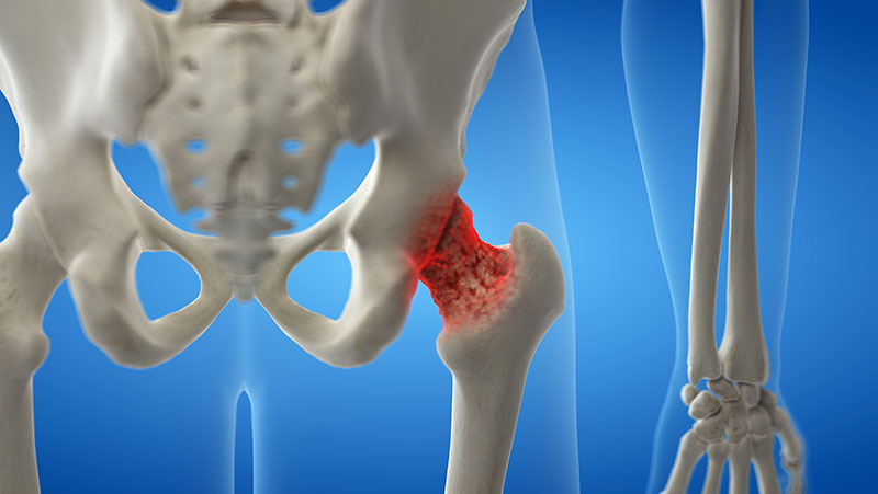
We believe Movement is Life!

Frozen Shoulder? New Research Indicates Dry Needling is Effective!

Here is the abstract from a recent journal article highlighting the results of the study.  Take home message: Frozen shoulder aka adhesive capsulitis is classically considered a “joint issue”.  Dry Needling aims to influence mainly “muscle dysfunction”.  What “joint issues” do you have that needling can help???

Trigger Point Dry Needling as an Adjunct Treatment for a Patient With Adhesive Capsulitis of the Shoulder

Authors: Derek Clewley, PT, DPT, OCS, FAAOMPT, Timothy W. Flynn, PT, PhD, OCS, FAAOMPT, Shane Koppenhaver, PT, PhD, OCS, FAAOMPT

Published: Journal of Orthopaedic & Sports Physical Therapy, 0, Volume: Early Access Issue: Early Access Pages: 1-32 doi:10.2519/jospt.2014.4915

Background: Prognosis for adhesive capsulitis has been described as self-limiting and can persist for 1-3 years. Conservative treatment including physical therapy is commonly advised.

Case Description: The patient was a 54 year old female with primary symptoms of shoulder pain and loss of motion consistent with adhesive capsulitis. Manual physical therapy intervention initially consisted of joint mobilizations of the shoulder region and thrust manipulation of the cervicothoracic region. Although manual techniques seemed to cause some early functional improvement, continued progression was limited by pain. Subsequent examination identified trigger points in the upper trapezius, levator scapula, deltoid and infraspinatus muscles that were treated with dry needling to decrease pain and allow for higher grades of manual intervention.

Outcomes: The patient was treated for a total of 13 visits over a 6 weeks period. After trigger point dry needling was introduced on the third visit, improvements in pain-free shoulder range of motion and functional outcome measures, including SPADI and QuickDASH, exceeded the minimal clinically important difference after 2 treatment sessions. At discharge the patient had achieved significant improvements in shoulder range of motion in all planes and outcome measures were significantly improved.

Discussion:This case report describes the clinical reasoning behind the use of trigger point dry needling in the treatment of a patient with adhesive capsulitis. The rapid improvement seen in this patient following the initiation of dry needling to the upper trapezius, levator scapula, deltoid and infraspinatus muscles suggests that surrounding muscles may be a significant source of pain in this condition.

Shock-wave therapy improved outcome with plantar fasciitis: a meta-analysis of randomized controlled trials

Abstract

Background: Shock-wave therapy (SWT) has been widely applied and proven to be an effective treatment in ameliorating symptoms of plantar fasciitis (PF). Ultrasound therapy (UT) is another common treatment of PF, and several researches have documented its advantages when compared to corticosteroid injection. Despite this, few studies have focused on comparing the use of SWT and UT in the treatment of PF. The purpose of our meta-analysis is to evaluate whether SWT is better than UT in managing PF, both in terms of ameliorating pain and improving functionality.

Methods: A systematic search of the literature was conducted to identify relevant articles that were published in Pubmed, Medline, Embase, the Cochrane Library, SpringerLink, Clinical Trials.gov and OVID from the databases’ inception to October 2018. All studies comparing the efficacy of SWT and UT in terms of pain levels and functionality improvement were included. Data on the two primary outcomes were collected and analyzed using the Review Manager 5.3.

Results: Five studies were included in the current meta-analysis. A significant difference in VAS score (MD = – 13.14, Cl – 14.07 to – 12.75 P < 0.00001, I2 = 100%) was noted between the SWT group and the UT group. No significant difference was seen in the AOFAS (MD = 3.19, Cl – 1.72 to 8.10 P = 0.20, I2 = 100%); FFI or PFPS score was not found significant difference either (SMD = – 1.17, Cl – 4.45 to 2.10 P = 0.48, I2 = 96%).

Conclusions: The results from this meta-analysis highlight the effectiveness of both SWT and UT in the treatment of PF. Although inter-group differences were not significant, the VAS score was better improved in the SWT group, suggesting that SWT may be a superior alternative for the treatment of PF.

3D Revolutionizes Gait Analysis

Since Aristotle’s text, “On the Gait of Animals,” human beings have been preoccupied with locomotion. While the philosopher writes of watching people’s shadows against a wall to observe gait patterns, today’s experts rely on more technical methods.

With the advent of 3D technology, gait analysis took a leap forward. What’s more, the honing of these systems over the years may have finally made them available to Joe and Jane Runner. The accuracy of the 3D systems are supported by both scientific and anecdotal evidence, making them perhaps the next big thing when it comes to treating and preventing running injuries.

3D Technology for the Masses

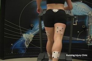
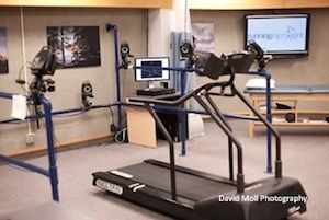
The use of 3D technology for gait analysis dates back to 1990. At the University of Calgary, Assistant Professor Dr. Reed Ferber, also the founder of the Running Injury Clinic in Calgary, uses a 3D system with eight cameras to gather data and images while research participants run on the treadmill. “I’ll tell you the exact degree your foot is overpronating. We can see if your lower leg is rotating appropriately with your foot and how much torsional force is being created at your knee,” explains Ferber.

Dr. Irene Davis, professor of physical therapy at the University of Delaware, utilizes a similar six-camera system in her Motion Analysis Laboratory. “In the lab, we measure mechanics in 3D and provide the runner with real time feedback on those mechanics,” says Dr. Davis. She argues, however, that offering this technology in the clinical setting may not be realistic. “At the end of the day, clinics aren’t going to have the manpower or financial resources for these expensive systems,” she says.

Dr. Ferber contends that the cost of such equipment has dropped dramatically over the past few years and that his system is real-world ready. “With respect to training, our software is turn-key in its use. We’re trying to make it relatively easy for the clinician to use this sophisticated technology to understand the root cause of an injury,” he explains.

He began putting the system to the test this April at a clinic in downtown Calgary, offering it to the general public for the first time. With the use of the 3D gait analysis system, he can observe runners’ overall biomechanics and gather data on the their flexibility, strength and alignment. With those four points of reference, he runs the numbers against two-and-a-half years worth of biomechanical profiles in his lab’s database and prints off a report to show patients the potential root cause of their injuries and what they might do to remedy them.

Dr. Ferber also offers healthy patients information on how to prevent injuries. “Based on those four pieces of the puzzle, we can say: You have a 63 percent chance of getting injured.” Dr. Ferber’s job is to help the runner understand how to lower their injury risk score. “We want to change the way runners think about their bodies and how they can reduce their risk,” he says. “We are making scientific decisions about what a runner needs to work on; we’re not taking a shot in the dark, we’re not guessing.”

Both Dr. Davis and Dr. Ferber agree that for a highly trained biomechanist, 2D high-speed cameras can also be accurate. “There’s nothing that takes the place of experience,” says Dr. Davis. “As you see more patients, you gradually develop your evaluation skills. With time, you begin to see specific mechanics associated with particular injuries.” While Dr. Ferber concurs, he believes that many of the people doing gait analyses simply don’t have the knowledge to be able to be able to provide precise feedback. He says that the 3D gait analysis system can offer this regardless of the operator’s background.

Plan of Action

While the cutting-edge gait analysis system will be of interest to techies, runners simply want to know what they can do to get healthy and resume running. Upon completing a gait analysis, Dr. Ferber and Dr. Davis offer differing methods of rehabilitation and injury prevention. While Dr. Ferber tends to use strength and flexibility prescriptions for his patients, Dr. Davis is partial to gait retrainment.

ric-clinic

Burning at each end of the candle, both have found success in their approaches. “I’m going to change a runner’s mechanics by getting them stronger and more flexible. Within six weeks, 90 percent of my runners are injury free,” says Dr. Ferber (results will be published soon in the Journal of Athletic Training).

Dr. Davis explains her research: “Our data suggests that you can alter the gaits of runners who are at risk for injury.” To do this she uses a treadmill and a mirror, giving the runner visual feedback on form and cadence.

Regardless of the rehabilitation protocol, gait analysis is becoming an increasingly important part of preventing and treating running injuries. Having seen success in his Calgary Clinic, Dr. Ferber already has plans to bring 3D gait analysis to Alberta, British Columbia and beyond. There is much anticipation for how this technology will affect the way we diagnose and treat running injuries. Aristotle would surely have been impressed.

The Importance of Gait Analysis

The equipment and methodology used for gait analysis has progressed substantially in recent years. The advances in how a person’s gait is evaluated, and the tools used to correct and restore normal gait, allow those who have suffered an injury affecting their ability to walk or run to be diagnosed more efficiently and accurately. Once diagnosed, a treatment plan for gait problems is created—usually by an experienced clinician—and is specifically designed to focus on the affected limbs with the goal of returning the patient to his or her highest level of functioning in the shortest amount of time.

Hope for Patients

Long gone are the days in which observation was the main diagnostic tool used by clinicians, and outdated methods of treatment have been replaced with state-of-the-art technology that’s changing lives. Accident victims, injured athletes, and those with central nervous and musculoskeletal system diseases and ailments all benefit from gait analysis. Regardless of gender, age, or degree of functioning, patients all over the country are benefitting from these technological advances, and this is an exciting, hopeful time for physical therapists and patients.

If you’re a physical therapist, working with injured athletes, people who have suffered limb injuries in accidents, or people diagnosed with multiple sclerosis or some other debilitating disease that affects muscle movement and control is more rewarding than ever before thanks to advances in gait analysis.

Clinical Gait Analysis

The term “clinical gait analysis” is the process by which information is gathered to fully understand the cause of gait abnormalities and to create a treatment plan best suited for the patient’s individual limitations and mobility goals. A variety of technology and methods are used for gait analysis, including, but not limited to the following:

  • Comprehensive physical examination
  • Motion and muscle assessment
  • Computer-interfaced video cameras used to measure motion (equipped with motion-analysis software)
  • Electrodes placed on the skin to monitor muscle activity
  • Force platforms utilized to measure force and torque of the patient to the ground
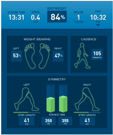
  • AlterG® Anti-Gravity Treadmill™ with Stride Smart

Physical therapists are using advanced gait analysis systems and video motion software to treat deficiencies in patients’ limbs and other areas of the body.  After just a few minutes of walking or running on a specially designed treadmill equipped with high-speed cameras, physical therapists are able to capture a patient’s movement while walking or running; this provides real time data to the physical therapists, so they can assess and treat a variety of gait-related injuries, regardless of the severity of injury and lack of mobility of the patient.

The instant feedback of some gait analysis technology allows physical therapists to more quickly create a treatment program that will help restore limb function to their patients with the goal of a shorter recovery time and a faster return to normal (or even improved) functioning. By using data to analyze how our bodies react to the movements they make and by evaluating the limbs’ reaction to force placed on them, physical therapists are able to teach their patients how to properly walk and run and advise patients as to what they can do to avoid—or, at least, minimize the chances of future injuries.

When it comes to gait analysis and equipment that monitors a patient’s progress, the diagnostic tools available today far exceed those available even just a few years ago. Stay tuned to the AlterG blog for information about how video is used to analyze, monitor and improve a person’s gait. Recording someone walking or running provides documentation of movements which will allow a physical therapist the opportunity to assess the fluidity and smoothness of a patient’s gait. Video tapes allow for close-up views of specific motions while walking and running, and when video is put in slow motion, the patient’s walking pattern is greatly enhanced making it much easier for a physical therapist to evaluate the movement of a patient.

Physical Therapy Guide to Osteoporosis

Osteoporosis is a common disease that causes a thinning and weakening of the bones. It can affect people of any age. Women have the greatest risk of developing the disease, although it also occurs in men. Osteoporosis affects 55% of Americans aged 50 or older; one-half of women and a quarter of men will fracture a bone as a result of low bone density (osteopenia) or osteoporosis. Thin bones are the cause of 1.5 million fractures per year in the United States; hip fractures alone result in 300,000 hospitalizations. It is important to diagnosis low bone density or osteoporosis early so that steps can be taken to rebuild bone strength and lessen the risk of fracture.

What is Osteoporosis?

Osteoporosis is a bone disease characterized by low bone density (thickness of the bone), decreased bone strength, and a change in the bone structure, which can lead to an increased risk of fracture. The normal bone structure becomes thinned out and porous with poor nutrition, aging, or when osteoporosis develops, lessening the ability of the bone to withstand the typical forces that are applied in everyday living. Fractures from low bone density and osteoporosis can be serious, causing pain and affecting quality of life.

Bone is living tissue. Normally, one type of cell removes bone and another type of cell adds bone in a balanced, ongoing process. In osteoporosis, bones weaken when not enough new bone is formed and/or too much bone is lost. This imbalance commonly begins in women during the first 5 years of menopause. However, it can also occur in men and in children, often due to diseases that affect bone development, such as celiac disease, inflammatory bowel disease, rheumatoid arthritis, spina bifida, cystic fibrosis, or kidney disease. Some medicines, such as steroids, may increase the risk of developing osteoporosis. Athletes who are underweight during the time of peak bone development are also susceptible.

There are many factors that can cause a person to be at risk for developing osteoporosis. It is important to know your risks so that you can be diagnosed and proactive in your treatment.

Risk Factors for Osteoporosis

Noncontrollable risks

  • Female gender
  • Small frame
  • Advanced age
  • Hormone levels
  • Genetics
  • Predisposing medical conditions

Controllable risks

  • Cigarette smoking
  • Excessive alcohol intake
  • Inactive lifestyle
  • Excessive caffeine intake
  • Lack of weight-bearing exercise
  • Drugs (eg, steroids, heparin)
  • Poor health
  • Low weight
  • Calcium-poor diet
  • Low vitamin D levels

How Does It Feel?

Osteoporosis is a disease that can be “silent.” There may be no outward symptoms until a fracture occurs. If you are middle-aged or older, you may notice a loss of height or the appearance of a humpback. You may also begin to experience pain between your shoulder blades or above the crest of the pelvis.

People with low bone density may experience fractures in everyday situations that would not occur in persons with healthy bones, such as breaking a hip or a wrist with a fall from a standing height, breaking a rib when opening a window or when receiving a hug, or breaking an ankle after stepping off a curb. These are called fragility fractures and are a red flag for bone disease. Spinal compression fractures, particularly those in the upper back or thoracic spine (area between the neck and the lower back), are the most common fractures, followed by hip and wrist fractures.

How Is It Diagnosed?

If you are seeing a physical therapist for back pain or other rehabilitation issues, the therapist will review your medical, family, medication, exercise, dietary, and hormonal history, conduct a complete physical examination, and determine your risk factors for osteoporosis. The assessment may lead the physical therapist to recommend further testing.

Osteoporosis is best diagnosed through a quick and painless specialized X-ray called the DXA, which measures bone density. The results are reported using T-scores and Z-scores.

  • The T-score compares your score to that of healthy 30-year-old adults. If you have a T-score of -1 or less, you have a greater risk of having a fracture.
  • If the T-score is -2.5 or less you will receive the diagnosis of osteoporosis.
  • The Z-score compares your bone mineral density to those of the same sex, weight, and age. It is used for those whose bone mass has not yet peaked, premenopausal women, and men older than 50.

Other methods of measuring bone density include X-ray, ultrasound, and CT scan.

How Can a Physical Therapist Help?

Your physical therapist can develop a specific program based on your individual needs to help improve your overall bone health, keep your bones healthy, and help you avoid fracture. Your physical therapist may teach you:

  • Specific exercises to build bone or decrease the amount of bone loss
  • Proper posture to protect your spine from fracture
  • Proper alignment during activities of daily living
  • How to improve your balance so as to reduce your risk of falling
  • How to adjust your environment to protect your bone health

Healthy bone is built and maintained through a healthy lifestyle. Your physical therapist will teach you specific exercises to meet your particular needs.

The exercise component for bone building or slowing bone loss is very specific and similar for all ages. Bone grows when it is sufficiently and properly stressed, just as muscle grows when challenged by more than usual weight. Two types of exercise are optimal for bone health: weight-bearing and resistance.

It is best for a physical therapist to provide your individual bone-building prescription to ensure that you are neither overexercising nor underexercising. Typically, exercises are performed 2 to 3 times a week as part of an overall fitness program.

Weight-bearing exercises

  • Dancing
  • Jogging (if your bone density is higher than -3.0)
  • Racquet sports
  • Heel drops
  • Stomping

Resistance exercises

  • Weight lifting in proper spine and lower-extremity alignment
  • Use of exercise bands
  • Gravity resistance (eg, push-ups, prone trunk extension with cushion to protect lowest ribs, single-leg heel raises, squats, lunges, sustained standing yoga poses in neutral spine position)
  • Exercises that reduce or stabilize kyphosis (hunchback)
  • Balance exercises

If you are diagnosed with osteoporosis or low bone density, your physical therapist will work with you to:

  • Build bone or lessen the amount of bone loss at areas most vulnerable to fracture through exercise—hip, spine, shoulder, arms.
  • Improve your dynamic balance to avoid falls.
  • Improve your posture.
  • Adjust your work and living environments to limit risk.
  • Help you avoid exercises and movements that may contribute to spinal fracture, including any type of sit-up or crunch, and excessive spinal or hip twisting.

Conservative treatment of a fracture includes bed rest and appropriate pain treatment. Your physical therapist will work with you to:

  • Decrease your pain through positioning and other pain-relieving modalities. Individualized physical therapist regimens can help reduce pain without the need for medications, such as opioids.
  • Provide appropriate external devices, such as bracing, to promote healing and improve posture.
  • Decrease your risk of a fall, strengthen your muscles, and improve your postural alignment.
  • Avoid exercises that involve too much forward or side bending or twisting.
  • Avoid water or endurance exercises, as they have been shown to negatively affect bone density.

If your pain lasts longer than 6 weeks following a spinal fracture, you can discuss surgical options, such as vertebroplasty or kyphoplasty, with your physical therapist, primary care physician, and surgeon.

Children and adolescents. Physical therapists can educate families and youth groups on proper exercise and posture, and about the need to move daily to build bone strength and prevent bone loss. Children with health issues such as spina bifida, diabetes, Crohn’s disease, and cerebral palsy are at a greater risk for bone disease and can particularly benefit from the guidance of a physical therapist. Proper physical conditioning is crucial for children and adolescents: the majority of bone is built during adolescence and peaks by the third decade of life.

Middle-aged and older adults. As people age, they may begin to notice postural, balance, and strength changes. Physical therapists work with middle-aged and older adults to:

  • Develop individualized exercise programs to promote bone growth or lessen bone loss
  • Improve dynamic balance to avoid falls
  • Improve posture
  • Improve the strength of back muscles
  • Improve hip strength and mobility

Can This Injury or Condition Be Prevented?

Osteoporosis can be prevented by building adequate bone density through childhood, adolescence, and early adulthood. Building strong bones requires an adequate intake of calcium and vitamin D, and regular exercise.

There are steps to take to improve bone health at any age. An active lifestyle that includes resistance and weight-bearing exercise is important to maintain healthy bone. It is also important to avoid habits that promote bone loss, such as smoking, excessive alcohol consumption, and an inadequate intake of calcium in your diet. Maintaining good body mechanics and posture also contribute to good bone health. We have no control over the genetic tendencies we have inherited, but we can choose to manage osteoporosis through proper medication, diet, and appropriate exercise.

As with any health issue, an overall healthy lifestyle is important for staying well.

Real Life Experiences

Anna is a 69-year-old retired legal secretary. She has enjoyed her early years of retirement, taking long walks in beautiful settings across the United States. Two years into her retirement, however, she began having knee pain during some of her walks, which gradually grew worse. Last year, she had a total knee replacement due to arthritis. She now walks with a cane because of chronic knee and ankle pain, and has experienced a loss of balance. She also has developed a rounded upper back, and low back pain. She seeks the help of a physical therapist.

Anna’s physical therapist performs an assessment that includes a medical review for osteoporosis risk factors and for other health issues. He evaluates her range of motion and strength, testing her arms, legs, and trunk—especially her upper back. He tests the flexibility of her spine and her balance, her walking ability, and her risk of falling. Anna’s walking style is uneven and she leans heavily on her cane. A DXA scan reveals that Anna has lost bone density in her spine and both hips. A vertebral fracture assessment X-ray shows that she has painless compression fractures of her spine. Her physical therapist diagnoses osteoporosis of the spine.

Anna first works with her physical therapist to improve her posture and knee function through flexibility and strengthening exercises, so she can walk more normally while working on her balance to lower her fall risk. She tells him her main goal is to be able to take walks in the park again.

Anna’s physical therapist teaches her safe trunk movement to avoid spinal fracture. Anna agrees to wear a dynamic trunk brace 2 hours a day to help make her posture more upright. She practices weight-bearing exercises with considerations for her arthritis, and learns resistive strengthening exercises for her spine and hip. Anna’s physical therapist designs a gentle home-exercise program for her as well.

By her last visit, the flexibility and strength of Anna’s trunk and legs and her tolerance of physical activity have improved. The quality of her walking and dynamic balance are measurably improved, and her risk of falling has decreased. Anna feels much more confident about managing her condition.

Just this past week, Anna joined a therapeutic senior walking group that meets at the local botanic garden twice a week. She is thrilled to be enjoying gentle walks in nature again, and looks forward to coordinating other activities with her new group of friends!

This story was based on a real-life case. Your case may be different. Your physical therapist will tailor a treatment program to your specific case.

What Kind of Physical Therapist Do I Need?

All physical therapists are prepared through education and experience to treat those with osteoporosis. However, if you have a diagnosis of osteoporosis or low bone density, you may want to consider:

  • A physical therapist who is a board-certified clinical specialist or who completed a residency or fellowship in orthopedic physical therapy or geriatric physical therapy. This physical therapist has advanced knowledge, experience, and skills that may apply to your condition.
  • A physical therapist who specializes in the treatment of osteoporosis.

You can find physical therapists with these and other credentials by using Find a PT, the online tool by the American Physical Therapy Association that can help you search for physical therapists with specific clinical expertise in your geographic area.

General tips when you’re looking for a physical therapist (or any other health care provider):

  • Get recommendations from family and friends or from other health care providers.
  • When you contact a physical therapy clinic for an appointment, ask about the physical therapists’ experience in helping people who have osteoporosis.

Physical Therapy Exercises After Hamstring Strain

If you have a hamstring strain or tear, you may benefit from the skilled services of a physical therapist to help you recover. Your physical therapist can assess your condition and prescribe treatments and exercises to help decrease your pain and improve your overall mobility.1

During physical therapy for a hamstring strain, your PT may use various modalities to help increase circulation, improve the way your muscles contract, and decrease pain.2 While these treatments may be beneficial, they should not be the only treatment you get for your hamstring condition.

Exercise is the most important component of your hamstring tear rehabilitation.3 But which exercises are best after a hamstring strain, and are there exercises to help you get back to your normal activity? Are there exercises to possibly prevent future problems with your hamstring? There are.

Your physical therapist can help determine the best exercises for your condition. This list is a sample progression of exercises that your PT may give to you during your rehab after a hamstring strain.

The exercise program starts out slow with some gentle stretching and range of motion (ROM) exercises, and it progresses in intensity until your hamstrings (and other neighboring muscles) are able to handle the high loads and stresses that are typically placed upon them.

Remember to check in with your doctor or physical therapist before starting this, or any other, exercise program for your hamstring strain rehab. Also, if any exercise causes lasting pain, stop it immediately and see your doctor.

Ready? Let’s get started.

1. Hamstring Stretches

A man stretching his hamstrings in the Gym
Gary Burchell / Getty Images

Imagine a big clump of balled up slop at the site where your hamstring muscle was torn. That tissue is collagen and is called scar tissue. It is the product of the normal healing process after a hamstring strain. The best way to remodel this tissue is with gentle, progressive stretching.4 (Your PT may also choose to do scar tissue massage with you to help get rid of that lump of tissue near your injury site.)

There are various ways to start to stretch your hamstrings after a hamstring strain. Start slowly and gently increase the intensity of each stretch over the course of four to six weeks.

Different hamstring stretches that your PT may prescribe for you may include:

  • The hurdler stretch5
  • The towel hamstring stretch
  • The amazing standing hamstring stretch

Each stretch can be held for 15 to 30 seconds, and you can do three to five repetitions. Hamstring stretches should be continued several weeks during your rehab, and you may wish to keep flexible by stretching your hammies several times a week even after you are fully healed.

2. Hamstring Strengthening Exercises

After performing a ball bridge, slowly bend your knees and allow the ball to roll towards your buttocks. Brett Sears, PT, 2011

Another part of your rehab should be focused on hamstring strengthening.2 Your PT can show you the best exercises to strengthen your hamstrings. Some ideas may include:

  • Prone knee curls (start with only gravity as your resistance, and then make things more challenging by adding cuff weights)
  • Standing hamstring curls6
  • Ball bridges with knee flexion
  • Seated hamstring curls with a resistance band
  • Good morning exercises
  • The Nordic eccentric hamstring curl7

Remember, start out slowly and then progress by increasing the number of repetitions of each exercise or by adding resistance with cuff weights or resistance bands.

Make sure you perform each strengthening exercise slowly and focus on the eccentric portion of the contraction. Eccentric contractions occur as your hamstring is lengthening.8 So, if you are doing a hamstring curl, controlling the motion slowly as your knee is straightening is the eccentric portion of the contraction. Some studies indicate that eccentric contractions may have a protective effect against hamstring injuries.

3. Calf Strengthening Exercises

Photo of a man running up the stairs.
The Alfredson Protocol can help treat your Achilles tendonitis. DaveLongMedia/ E+/ Getty Images

Remember, your hamstrings cross your knee joint in the back. Guess what? Your calf muscles cross there too, so don’t ignore them while rehabbing a hamstring injury. Your calf and hamstring work together to help support your knee, so performing calf strengthening exercises can help support your entire lower extremity.

Exercises to strengthen your calves may include:

  • Calf raises9
  • Theraband strengthening exercises
  • Alfredson protocol for Achilles strength10

Each different exercise should be done for 10 to 15 repetitions several times per week. Stop if any calf exercise starts to increase pain in your injured hamstring.

4. Hip and Quad Stregnthening Exercises

Woman running with hip muscles exposed
Henning Dalhoff / Getty Images

Your hip muscles, like the gluteus medius, help control the position of your leg as you are walking and running. If your hips are weak, your lower leg may rotate inwards and place excessive stress on your knee and the muscles around it. Keeping your hips strong can help alleviate this excessive stress on your hamstring muscles, which may help protect them from further injury after a hamstring strain.11

Straight leg raises are a great way to start your hip strengthening exercises. Once your hips get stronger and your hamstring has healed, you can start more advanced hip strengthening, like hip hikers.

Your quadriceps muscles are on the front of your thigh, just opposite your hammies. Keeping these muscles strong can help support your entire lower extremity and create balance between all the muscles of your leg. Short arc quad exercises or mini squats can help keep your quads working well while you rehab your hamstrings.

Exercises for your hips and quads should be done for 10 to 15 repetitions, 3 to 4 times per week.

5. Abdominal and Core Stability

Photo of a woman doing a plank with a toddler on her back.
You can use just about anything at home to do your PT exercises. Getty Images

Your abdominal and core muscles attach to the top of your pelvis, and your hamstrings originate from the bottom of your pelvis. Therefore, keeping your core strong can affect pelvic position, and a pelvis that is not in optimal position may place increased stress and strain on your hamstring muscles. It makes sense that your physical therapist may have you work on core stability exercises as part of your hamstring strain exercise program. Research indicates that hamstring rehab programs that include core exercises may yield effective results.12

Abdominal and core strengthening exercises that your PT may prescribe include:

  • The pelvic tilt
  • Bridges13
  • Stability ball bridges
  • Planks14
  • Quadruped exercises

Remember to start with an easy exercise that places minimal stress on your hamstrings, and progress to more challenging exercises as your hamstring heals and you are able to tolerate more strain on your muscles.


6. Balance and Proprioception Exercises

Photo of a wobble board.
A wobble board can provide an unsteady surface on which to perform balance exercises. Rollover/Getty Images

Your physical therapist may have you work on balance and proprioception exercises as part of your hamstring strain rehab program. Poor balance and instability can make your leg muscles, like your hamstrings, work extra hard during high-intensity athletics like running or jumping.15 This may place excessive stress on your hamstrings, leading to a strain.

Balance exercises may include:

One important caveat with balance exercises: be safe. To effectively improve your balance, you must create situations that challenge your balance, and this may place you in a situation where you can fall. Be sure you remain safe while performing your balance exercises by keeping something nearby that you can hold onto to stabilize yourself.

Your physical therapist can show you the best exercises to do to improve your balance during your hamstring rehab.


7. Plyometric and Return to Sport Exercises

Photo of an athlete doing a box jump.
Plyometric training may be a part of your ankle fracture rehab. John Fredele/Getty Images

As your hamstring strain physical therapy program progresses, your PT may have you work on high-intensity jumping exercises, called plyometrics. Exercises may include:

All of these exercises have one thing in common: they place high amounts of stress on your lower extremity muscles and joints.18 They should be done under the close supervision of your physical therapist and are reserved for the latter stages of your hamstring strain rehab when significant healing has taken place.

Your physical therapist can also perform functional mobility testing to determine when (and if) it is safe for you to return to high-intensity athletics. These tests are designed to challenge your leg muscles, like the hamstrings, and can help your PT determine if you can get back to sports.

Once your hamstring has healed and you are ready to move on from the physical therapy clinic and back to normal activity, your PT can work with you to devise a home exercise program to help you maintain optimal flexibility, strength, balance, and jumping ability to help you minimize your risk of future hamstring strains.

Keep in mind that everyone is different, and everybody heals at different rates. Plus, every injury is not the same, and your hamstring recovery may be quick, or it may take a bit longer than you would like. The best way to understand what exercises you should do for your hamstring rehab is to work closely with your doctor and physical therapist.

Be patient with your body during your recovery, and you’ll get back to doing the things you were doing prior to your injury. Your PT can ensure that you do the correct exercises, at the correct time, to safely get you back to your optimal level of activity.

A Word From Verywell

If you have had a hamstring strain, your PT will likely progress you through exercises, similar to this program, to help you fully recover. Check in with your physical therapist to start on your own personalized rehab program for your hamstring strain so you can get back to optimal functional mobility quickly and safely.